
Case reveals tooth in patient’s nasal cavity
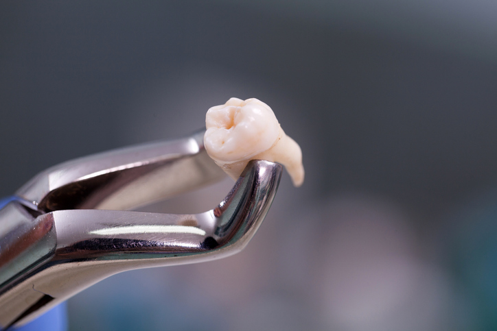
While a rare clinical entity, intranasal ectopic teeth can be accompanied by symptoms that make it difficult to identify — including obstruction, rhinorrhea and sinusitis — as well as recurring sinonasal complications.
In a case report published in Radiology Case Reports, researchers detailed a 45-year-old male patient who was experiencing right-sided nasal discharge with a foul smell and the sporadic occurrence of epistaxis. The patient’s symptoms had been ongoing for at least six months, and upon examination with nasal endoscopy, the researchers observed a hardened mass with calcified deposits in the right nasal cavity.
Following computed tomography scans, the researchers discovered that the mass contained hyperdense material, a central pulp cavity and a rhinolith — representing an intranasal ectopic tooth. Treatment with endoscopic transnasal extraction successfully removed the ectopic tooth, and the patient experienced no symptoms or complications during recovery.
The findings suggested that clinicians should consider intranasal ectopic teeth among patients presenting with ongoing unilateral nasal symptoms and highlighted imaging and minimally invasive treatment strategies that may be effective in this patient population.
Read more: Radiology Case Reports
The article presented here is intended to inform you about the broader media perspective on dentistry, regardless of its alignment with the ADA's stance. It is important to note that publication of an article does not imply the ADA's endorsement, agreement, or promotion of its content.